Working Time
- Mon - Sat: 9 AM - 4 PM
Sun : 9 AM - 2 PM
Contact Info
-
Phone: 9990507691, 9811892896
- srivastavamri@gmail.com
Ask the Experts
ULTRASOUND SCAN

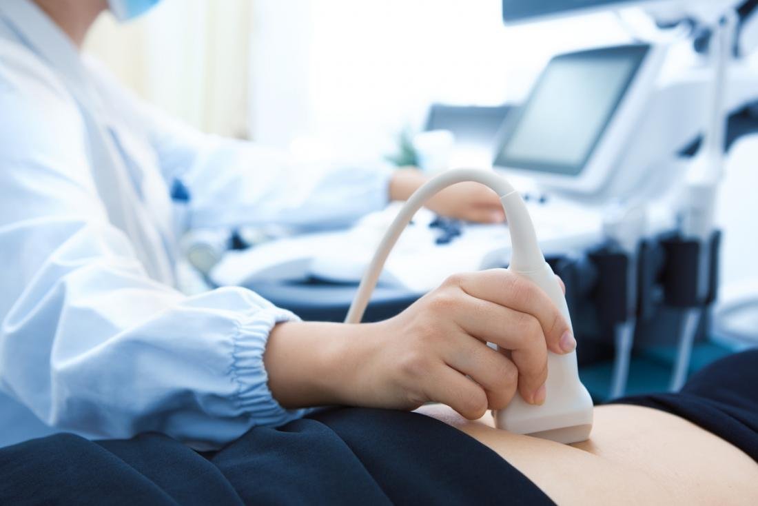
An ultrasound scan or sonography is a process that uses sound waves that are in high frequency to generate an image of parts inside of the body instead of radiation. It is used to monitor or evaluate the unborn child and diagnose the problems in the liver, heart, kidney, or abdomen. It helps the doctor to see the problems with vessels, tissues, and organs without making any incision. It may also help in performing certain types of biopsy/FNAC. It is often used to check the progress of a pregnancy.
3D & 4D Ultrasound:
3D Ultrasound scans show the three-dimension pictures of the baby. 4D scans are also similar to 3D ultrasound scans; the only difference is that in 4D there are 3-spatial dimensions and 1-time dimension. Couples who are disappointed when they see that their scan is a grey and blurry outline can go for 3D and 4D scans.
In this, you can see your baby's skin, your baby’s mouth and nose shape, and other activities that he/she doing inside your womb. These scans are also safe as 2D scans because pictures are made up of sections of 2D images. These scans are done only by doctor recommendation because it takes more time to create 3D scans, so the doctor should allow the patient for these scans. These scans are also useful to look at the heart and other internal organs.
WHY IS IT DONE?
Most people think that ultrasound is connected with pregnancy only. The scan provided the first view of the unborn babies. But, it is used to diagnose other problems also like if you have any swelling, pain, infection or other symptoms, it will diagnose through ultrasound scan by an internal view of your organs. It can provide the view of:
- Infant’s brain
- Gallbladder
- Eyes
- Kidneys
- Liver
- Ovaries
- Pancreas
- Thyroid
- Testicles
- Uterus
- Blood vessels
HOW IS IT DONE?
Before going to the exam, you may need to wear the hospital gown; sometimes it wouldn’t need to change. After that, you will likely to lying down on a table and exposed that section of the body that needs to be scanned. Later our expert will apply a special lubricating gel to the scanned part of the body. A small probe is used called transducer that helps to transmit the sound waves and also prevents the friction with the help of lubricating gel.
That sound waves travel from the transducer through the gel into the body and collects the sounds that bounce back into a computer. The expert may ask you to change your position to have better access. After the scan, the gel will be wiped off from your skin. The procedure may last less than 30 minutes according to the part of the body being examined.
Tips before going to the procedure:
- If your abdomen is being examined, you may be told to fast for approx. 8-12 hours before going to the examination.
- If your liver, gallbladder and pancreas are being examined, you may be told to eat a fat-free meal.
- For other examinations, you may be told to drink a lot of water till your bladder is full and you need to hold your urine to get better visualization.

