

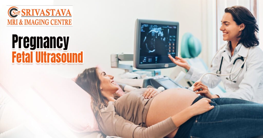
High-frequency sound waves are transmitted through the belly during prenatal ultrasound or ultrasonography using a device known as a transducer. The sound waves are captured and converted into baby images for video or photography. An ultrasound during pregnancy can display pictures of the ovaries, placenta, and amniotic sac. An ultrasound might be performed earlier in your pregnancy to determine:
1. Heartbeat and movement of its body, arms, and legs
2. Presence of more than one fetus
3. Due date or gestational age (the age of the fetus)
4. Later in pregnancy, an ultrasound might be used to determine:
5. Fetal well-being
6. Placenta location
7. Amount of amniotic fluid around the baby
8. Position of the baby
9. Baby’s expected weight
10. Major anatomical abnormalities or birth defects
There are different types of ultrasound performed during your pregnancy journey:
Dating and Viability Ultrasound
This is the very first 7 weeks of pregnancy ultrasound and is used to confirm pregnancy status in women.
Transvaginal ultrasound
It is 12-week pregnant ultrasound used during the early stages of pregnancy. A needle is inserted into your vagina and used to image clear images.
Nuchal Translucency Ultrasound
This is a more detailed ultrasound that displays the width, length, and depth of the fetus and images of its organs. This is a 14 – 18 weeks pregnant ultrasound. Nuchal translucency (NT) test to check for Down syndrome, heart defects, or other chromosomal abnormalities in the fetus. It uses a special probe and software and is available only in some hospitals.
Anatomy Scan
This is 18 and 20 weeks pregnant ultrasound in the second trimester. It is detailed pregnancy ultrasound. This is a 4D ultrasound’, this ultrasound creates a live video of the fetus. This dynamic video captures multiple images of the baby’s facial expressions, and movements, and also shows glimpses and shadows.
Fetus Echocardiography
This is used to diagnose fetal heart problems. This is similar to a traditional pregnancy ultrasound.
Transabdominal ultrasound
This sends waves to your lower abdomen using a converter and special gel. Images are painted in black and white to depict fetus development and abnormalities.
Doppler ultrasound
This is a type of ultrasound scan in pregnancy used to assess blood circulation in your baby’s heart. In this procedure, a thin amplifier is used to listen to your baby’s heartbeat.
If you wanted to make your pregnancy journey blissful, you are welcome to Srivastava MRI & Imagine center, the best-rated, state-of-the-art, premium, and exclusive Ultrasound Centre in Jasola. You may go another branch to enhance the joy of your parenthood with Ultrasound Centre in Mayur vihar.