Working Time
- Mon - Sat: 9 AM - 4 PM
Sun : 9 AM - 2 PM
Contact Info
-
Phone: 9990507691, 9811892896
- srivastavamri@gmail.com
Ask the Experts
MRI

One stop solution for all your diagnostic needs! / Complete Diagnostic solution!
WHAT IS MRI?
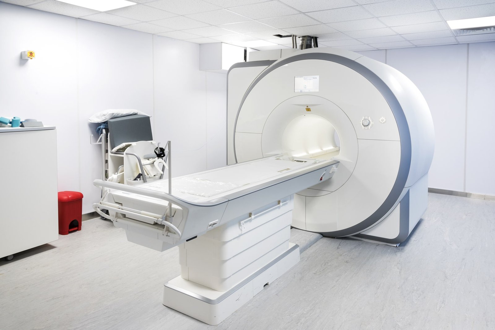
Magnetic Resonance Imaging (MRI) is one of the most advanced diagnostic commodities available to mankind. It is a test that uses powerful magnets, radio waves, and a computer to make detailed pictures inside your body. MRI marks a giant leap in imaging technology by doing away with harmful ionizing radiations and producing digital images that allow easy accessibility and detailed study.
This test is used to diagnose or to see how well you have responded to treatment. Unlike X-rays and computed tomography scans (CT scans), MRI doesn’t use the damaging ionizing radiation of X-rays.
WHY IS IT DONE?
An MRI protocol is highly sensitive and provides a comprehensive array of images that help a doctor diagnose the injury, tumor, bleeding, infection or disease which may be missed under similar X-ray or CT scans protocols. It can also be used to monitor patients undergoing treatment. Dedicated MRI coils are available for the brain, spinal cord, heart, blood vessels, bones and joints, liver, kidney uterus, and other internal organs. It is an entirely painless and non-invasive procedure that is rapidly changing the field of diagnostic and imaging.
HOW IS IT SCAN DONE?
At Srivastava MRI and Imaging Centre, we provide a scanning facility for the complete body as well as dedicated coils for in-depth study of isolated organs and body structures. An average scanning protocol takes about 20-40 minutes during which the patients lie down calmly on a motorized table while the scanner takes multiple images of the region. These images are then processed by a dedicated computer for clarity and our team of well-qualified radiologist reviews these scans to uncover the underlying aetiology.
Let’s see how the procedure is performed:
- The patient will be positioned on the movable exam table. Straps and bolsters may be used to help the patient to stay still and maintain the position.
- A device which contains coils capable of sending and receiving radio waves may be placed around or next to the area of the body being scanned.
- MRI exams generally include multiple sequences. If a contrast material is used, a technologist will insert an IV line into vein in your hand that will be used to inject contrast media.
- You will be placed on the motorized bed that allows you into the magnet of the MRI unit. The technologist will perform the scan while working at a computer outside the magnetic room.
- If a contrast media is used during the scan, it will be injected intravenous after an initial series of scans. Once done, your IV line is removed after the scan is over.
- When the exam is complete, you may be asked to wait while the radiologist or technologist checks the images in case more are needed.
- Depending on the type of exam the entire exam is usually completed in 20-40 minutes.
Tips before going to the procedure :
- You have to wear a gown provided by the hospital or you may also be allowed to wear your own clothing, but it should be loose-fitting and has no metal fasteners.
- Leave all jewelry and other metallic accessories at home or remove them before going to the MRI scan because it may cause burns or become harmful projectiles within the MRI scanner room.
- The accessories include watches, credit card, jewellery, hearing aids, hairpins, metal zipper, and other metallic items such as removable dentures, mobile phones, knives, eyeglasses, pens, and many more.
- Some MRI exams use an injection of contrast media, so you may be asked if you have asthma or allergies to any drugs. MRI exams commonly use a contrast media called gadolinium.
- Tell the technologist or doctor if you have any serious health problems or recently had surgery.

